Clinical, Histopathologic, Immunohistochemical, and Electron Microscopic Findings in Cutaneous Monkeypox: A Multicenter Retrospective Case Series in Spain
December 2022
in “
Journal of the American Academy of Dermatology
”
TLDR Monkeypox skin lesions show full-thickness skin death and swollen skin cells, with the virus found in affected cells.
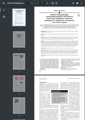
This retrospective study analyzed the histopathologic and immunohistochemical characteristics of cutaneous lesions in 20 patients with confirmed monkeypox infection. The most consistent histopathologic findings were full-thickness epidermal necrosis and keratinocytic ballooning, with some cases also showing involvement of the hair follicle's outer root sheath and sebaceous gland epithelium. Immunohistochemical analysis revealed strong positivity for Vaccinia virus in the cytoplasm of ballooned keratinocytes. Electron microscopy, performed in 4 cases, confirmed the presence of numerous monkeypox viral particles in affected keratinocytes. These results underscore the value of histopathologic and immunohistochemical examination of skin lesions in diagnosing monkeypox, despite the study's limitations, including its small sample size and the limited number of cases examined by electron microscopy.