Three-Dimensional Imaging of Tight Junction Network Across Multiple Layers of Human Epidermis by Array Tomography Using Backscattered Electron-Mode Scanning Electron Microscopy
August 2019
in “
Journal of Investigative Dermatology
”
epidermal turnover keratinocyte proliferation epidermal differentiation markers desmoplakin epidermal morphogenesis PPARs Ppara gene expression skin barrier integrity microbial diversity BP180 Dsg1 degradation tight junctions stratum granulosum skin turnover skin cell proliferation skin differentiation markers skin structure skin barrier skin microbes skin protein skin layers

TLDR The study found that tight junctions reach the top layer of the skin's stratum granulosum, not just the second top layer as previously thought.
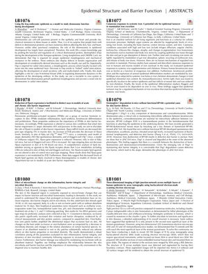
The document presents abstracts from various studies related to epidermal structure and barrier function. One study (LB1077) suggests that epidermal turnover may be a regulated mechanism of iron excretion, with keratinocyte proliferation and expression of epidermal differentiation markers being modulated by iron. Another study (LB1076) finds that desmoplakin is essential for proper epidermal morphogenesis and mechanical resistance in Xenopus embryos, with a role in radial intercalation. The study (LB1078) on PPARs shows that Ppara gene expression is reduced in models of chronic skin impairment but not in acute skin barrier impairment, with lipid metabolites likely involved in Ppara downregulation. Research (LB1080) on skin microclimate indicates that occlusion and over-hydration negatively affect skin barrier integrity and microbial diversity, but these effects can be mitigated by absorbent materials. Another study (LB1079) reports that dysfunction of BP180 leads to Dsg1 degradation and defective skin barrier, which may cause an atopic dermatitis-like phenotype. Lastly, the study (LB1081) aimed to resolve the controversy about the extent of tight junctions (TJs) in the epidermis by using array tomography with backscattered electron-mode scanning electron microscopy, revealing that TJs extend to the most superficial layer of the stratum granulosum, contrary to previous suggestions that they were only present in the second most superficial layer.


