TLDR  A man had a rare pigmented nodule on his scalp that developed from birthmarks.   
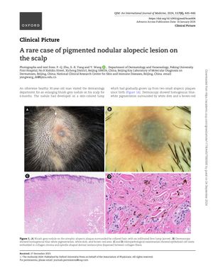
  A 30-year-old man presented with a bluish-grey nodule on his scalp, which had developed over 6 months from a skin-colored lump originating from two alopecic plaques present since birth. Dermoscopy revealed homogenous blue-white pigmentation, white dots, and a brown-red area. Histopathological examination identified epithelioid cell nests in collagen stroma and spindle-shaped dermal melanocytes among collagen fibers.
          
        4 citations
,
  October 2012   in “Expert Review of Dermatology”
           4 citations
,
  October 2012   in “Expert Review of Dermatology”    Dermoscopy greatly improves melanoma diagnosis and reduces unneeded surgeries.  
      May 2009   in “The American Journal of Dermatopathology”
           May 2009   in “The American Journal of Dermatopathology”    Mast cells play a significant role in hair loss conditions like male pattern hair loss and alopecia areata.  
      November 2019   in “Harper's Textbook of Pediatric Dermatology”
           November 2019   in “Harper's Textbook of Pediatric Dermatology”    The document is a detailed medical reference on skin and genetic disorders.  
      7 citations
,
  July 2019   in “Clinics in Dermatology”
           7 citations
,
  July 2019   in “Clinics in Dermatology”    Hair loss can indicate or worsen with systemic diseases, and treating the underlying condition is important.  
      December 2024   in “Archiv Euromedica”
           December 2024   in “Archiv Euromedica”    Trichoscopy is essential for diagnosing and treating autoimmune cicatricial alopecia early.