Phylloid Terminal Hair Nevus: A Unique Clinical Entity
August 2018
in “
Pediatric Dermatology
”
phylloid terminal hair nevus phylloid hypopigmentation terminal hairs phylloid hypomelanosis hypopigmented skin dilated follicular structures tuberous sclerosis mosaic trisomy 13q phylloid pigmentary mosaicism phylloid nevus hypopigmentation dark brown hairs hypomelanosis light skin patches follicular structures genetic alterations pigmentary mosaicism

TLDR A baby had a unique skin condition with a pale patch and surrounding dark hairs, not linked to other health issues.
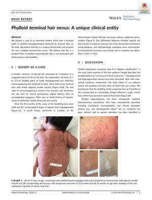
In 2018, a case was reported of an infant with a unique skin condition characterized by a localized patch of phylloid hypopigmentation bordered by terminal hairs on the back, which the authors proposed to call "phylloid terminal hair nevus." This condition was hypothesized to be a localized form of phylloid hypomelanosis without associated extracutaneous abnormalities. The 19-day-old female patient presented with a 3.1 by 2.3 cm patch of subtly hypopigmented skin surrounded by thick, dark brown terminal hairs. Over time, many bordering hairs were shed, and the central patches became more hypopigmented. A punch biopsy showed dilated follicular structures but no other abnormalities. The patient did not exhibit any signs of tuberous sclerosis or genetic alterations commonly associated with phylloid hypomelanosis, such as mosaic trisomy 13q. At 14 months, the patient was developing normally without extracutaneous disease. This case was distinguished from other forms of phylloid pigmentary mosaicism by its localized nature and the presence of terminal hairs, leading the authors to suggest it as a distinct clinical entity within the spectrum of phylloid pigmentary mosaicism.


