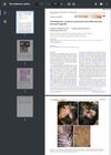
Lipidized Fibrous Histiocytoma: Differential Diagnosis from Juvenile Xanthogranuloma
January 2019
in “
Annals of Dermatology
”
TLDR The study concluded that careful examination is key to differentiate between lipidized fibrous histiocytoma and juvenile xanthogranuloma.
In 2018, a case study was conducted on a 33-year-old male who presented with an asymptomatic papule on his left forearm, which had been present for 6 months. The papule was diagnosed as lipidized fibrous histiocytoma (FH), a rare subtype of FH that accounts for 2.1% of FH variants. Lipidized FH is often misdiagnosed as juvenile xanthogranuloma (XG), a common benign skin tumor that affects children's head and neck. The study emphasized the importance of histology and immunohistological techniques in distinguishing between these two diseases when clinical diagnosis is challenging. The patient's tumor was completely removed by punch excision, and the diagnosis was confirmed through histopathological examination and immunohistochemical staining.