Diagnosis: Alopecia Areata or Not?
March 1999
in “
Seminars in Cutaneous Medicine and Surgery
”
alopecia areata trichotillomania telogen effluvium androgenetic alopecia scalp biopsy pigment casts follicle phases peribulbar lymphocytic infiltrates follicle miniaturization horizontal sectioning hair loss hair pulling disorder stress-related hair loss male pattern baldness biopsy hair follicle changes immune cells around hair follicles shrinkage of hair follicles

TLDR Scalp biopsies are essential for accurately diagnosing alopecia areata.
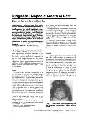
In 1999, Diane M. Hoss and Jane M. Grant-Kels highlighted the diagnostic challenges of alopecia areata and its differentiation from conditions like trichotillomania, telogen effluvium, and androgenetic alopecia. They presented four cases where scalp biopsies were crucial in making the correct diagnosis. In two cases involving adolescent females, the biopsies helped differentiate trichotillomania from alopecia areata by identifying pigment casts and changes in follicle phases. In the other two cases of adult women with diffuse hair loss, the biopsies distinguished diffuse alopecia areata from other conditions through the detection of peribulbar lymphocytic infiltrates and follicle miniaturization. The article underscored the significance of scalp biopsies, recommending horizontal sectioning of the biopsy specimen for a more informative result and referencing the "Color Atlas of Differential Diagnosis of Hair Loss" as a diagnostic guide.


