Reflection Confocal Microscope Characteristics of Rosacea and Related Observations
July 2022
in “
Skin research and technology
”
rosacea reflection confocal microscope skin CT hair follicles sebaceous units atrophied epidermis flattened epidermis spongy edema spinous layer hair follicle sebaceous gland units perifollicular abscesses proliferation of blood vessels dilation of blood vessels inflammatory cell infiltration Demodex structures Demodex mites

TLDR Skin CT can help diagnose rosacea by identifying specific skin features, but should be used with clinical signs to avoid misdiagnosis.
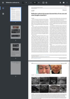
The study conducted by Hongyong Sun and colleagues used a reflection confocal microscope (also known as skin CT) to examine the skin of patients diagnosed with rosacea, a chronic inflammatory disease that affects facial blood vessels, nerves, hair follicles, and sebaceous units. The researchers identified several common skin CT features in these patients, including atrophied and flattened epidermis, spongy edema in the spinous layer, increased diameter of hair follicle sebaceous gland units, formation of perifollicular abscesses, proliferation and dilation of blood vessels around the hair follicle, and increased inflammatory cell infiltration around blood vessels and hair follicles. Some patients also showed colonization of Demodex structures in hair follicle sebaceous gland units. The researchers caution that these features should be used in conjunction with clinical manifestations to avoid misdiagnosis of rosacea as other diseases with similar skin CT features.


