 August 2018 in “Pediatric Dermatology”
 This case report describes a unique instance of an otherwise healthy infant with phylloid terminal hair nevus, a form of hypomelanosis without extracutaneous abnormalities.
 1 citations
,
November 2021 in “Biomedicines”
1 citations
,
November 2021 in “Biomedicines” This review elaborates on the concept of cutaneous mosaicism and its link to acneiform conditions, but it reports no new clinical results.
 2 citations
,
January 2019 in “Springer eBooks”
2 citations
,
January 2019 in “Springer eBooks” This review discusses segmental vitiligo as part of the vitiligo clinical spectrum and highlights its role as a model for studying repigmentation, but reports no new clinical results.
 November 2019 in “Harper's Textbook of Pediatric Dermatology”
 This index of Harper's Textbook of Pediatric Dermatology, Fourth Edition, provides no clinical results or new findings.
 February 2025 in “Indian Dermatology Online Journal”
 This study explored the dermatological phenomena associated with sports and clinical scenarios, detailing unique conditions like "Athlete's nodule," "Runner's toe," and "Swimmer's ear," along with their diagnostic features and management strategies, uncovering how sports activities inform dermatological terminology and practice.
January 2026 in “Annals of Dermatology” This review outlines evidence-based strategies for diagnosing pediatric hypopigmented disorders and emphasizes distinguishing vitiligo from self-limiting conditions through a systematic clinical approach; no new results are reported.
6 citations
,
February 2020 in “Journal of Cutaneous Pathology” This case report describes the histopathological features of nevus psiloliparus in an 11-year-old girl, noting "shadow" follicular units with loosely arranged collagen and reduced elastic fibers.
56 citations
,
October 2010 in “Pediatric Clinics of North America” This article discusses various types of epidermal nevi and associated syndromes but presents no new research findings.
6 citations
,
September 2014 in “Journal of the American Academy of Dermatology” In this report, researchers described a potential new variant of mucinous nevus, characterized by dilation of hair follicle pores and fibromucinous stroma in both the papillary and perifollicular dermis.
44 citations
,
January 1999 in “Dermatology” This article reviews different perspectives on nevus comedonicus, discussing its classification and potential associations with systemic findings, but it reports no new results.
1 citations
,
January 2019 in “Dermatology Online Journal” This case report describes a rare occurrence of nevus comedonicus on the scalp of a 19-year-old woman, highlighting its unusual location and classic appearance.
October 1967 in “Archives of Dermatology” This report discusses a case of an intradermal nevus in a 42-year-old woman and provides no clinical findings beyond a biopsy showing hyperkeratosis and cellular pigmentary changes.
September 2024 in “The Journal of Dermatology” In this study, researchers reported a rare case of nevus comedonicus with hair growth in a 26-year-old male, challenging prior reports that affected lesions typically lack the capability to develop terminal hair.
7 citations
,
January 2013 in “Ophthalmic plastic and reconstructive surgery” This case report describes a rare instance of late-onset, bilateral nevus comedonicus of the eyelids in a 79-year-old man, highlighting its clinical and histopathologic features.
January 2025 in “Indian Journal of Dermatopathology and Diagnostic Dermatology” In this histopathology study of nevus sebaceus, researchers observed distinct epidermal, sebaceous, follicular, and apocrine changes, including sebaceous hyperplasia and basaloid adnexal proliferation, which help differentiate it from similar conditions like basal cell carcinoma and trichoblastoma.
3 citations
,
March 1966 in “Archives of Dermatology” This study found that nevoid basal cell carcinomas originate in the epidermis and the upper part of the hair follicle, showing similarities to nonnevoid basal cell carcinomas.
April 2025 in “Indian Journal of Dermatology” In this case report, a 24-year-old male with a translucent nodule in the left nasolabial fold was diagnosed with a solitary fibrofolliculoma with cystic changes upon histopathology, emphasizing the rarity and potential for misdiagnosis of these benign cutaneous tumors.
2 citations
,
May 1991 in “PubMed” This article reviews naevus comedonicus, highlighting potential treatments like retinoic acid application and surgical intervention, but reports no new clinical findings.
21 citations
,
April 2000 in “Journal of Cutaneous Pathology” This case report describes a 22-year-old woman with a nevoid plaque termed localized follicular hamartoma, characterized by skin-colored papules and specific cellular and structural features.
61 citations
,
March 1966 in “Archives of Dermatology” This study found that nevoid basal cell carcinomas originate in the epidermis and upper hair follicles, resembling early nonnevoid basal cell carcinoma proliferations.
2 citations
,
April 2012 in “American Journal of Dermatopathology” This report describes two cases of nevus lipomatosus superficialis with the unusual feature of dilated hair follicles, emphasizing the need for precise histopathological diagnosis to differentiate from similar conditions.
 November 2017 in “International journal of research in dermatology”
 This case report describes a rare instance of nevus comedonicus with a multi-dermatomal unilateral pattern along the lines of Blaschko, treated unsuccessfully with topical tretinoin and oral retinoids.
12 citations
,
August 1988 in “Histopathology” This case report describes a giant pigmented tumor of the scalp in a 47-year-old woman and suggests a possible dual origin involving neural crest differentiation.
31 citations
,
January 2007 in “Journal of the American Academy of Dermatology” A rare skin growth was successfully removed without recurrence after one year.
1 citations
,
May 2007 in “Chinese Medical Journal” This case report describes a 24-year-old woman with a rare giant cerebriform pigmented nevus on the scalp, which was identified as a giant congenital intradermal nevus based on clinical and pathological findings.
5 citations
,
August 2000 in “Archives of Pathology & Laboratory Medicine” This case report describes an 89-year-old woman with a recurrent benign proliferating trichilemmal tumor on her back, highlighting its misdiagnosis as a sebaceous cyst and successful excision with free margins.
 March 2026 in “Scientific Journal”
 This case study reported a 15-year-old girl with a nevus sebaceus of Jadassohn, where a secondary neoplasm potentially developed into either pigmented trichoblastoma or pigmented basal cell carcinoma, highlighting the importance of early recognition and evaluation of such lesions for effective management.
19 citations
,
July 1994 in “International Journal of Dermatology” This case report describes a 9-year-old Korean boy with a calcified nodule on his earlobe, confirmed by histopathologic examination and Von Kossa staining.
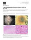
This study reported the case of a 30-year-old man with a bluish-grey scalp nodule that developed over a previously stable hairless plaque, revealing histopathological features consistent with epithelioid cell nests and spindle-shaped dermal melanocytes embedded in collagen.
38 citations
,
June 2005 in “International Journal of Dermatology” This case report describes a 28-year-old man with widespread pigmented basal cell carcinomas and recurrent infections that were challenging to control, associated with nevus comedonicus and a rudimentary toe.