Preparation of Mohs Micrographic Surgery Frozen Sections: Three New Pearls Leading to a Simplified, More Effective Process
May 2013
in “
Dermatologic Surgery
”
TLDR Three new techniques simplify and improve the preparation of tissue samples for skin cancer surgery.
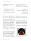
The document describes three new techniques to improve the preparation of frozen sections for Mohs Micrographic Surgery (MMS), which is crucial for visualizing surgical margins and minimizing artifacts. The first technique involves using a frozen glass slide to adhere the specimen, allowing for immediate visualization of all margins and the identification of air pockets. The second technique modifies the embedding mold to create a level surface for the specimen, which prevents "tip-lift errors" and allows for immediate sectioning without the need for rough cutting. The third technique uses colored optimal cutting temperature (OCT) medium to seal any crevices and indicate when an en face specimen is achieved during sectioning. These methods aim to simplify the process, increase efficiency, and ensure complete margin visualization. The document also briefly discusses the challenges of follicular unit extraction (FUE) in African-American hair for hair transplantation, but this is not the main focus of the summary.