Histopathologic Profile of Alopecia Areata in Indian Patients
January 2010
in “
International Journal of Trichology
”
TLDR The study found that the cause of alopecia areata can be identified through tissue analysis, and vertical sections are enough for diagnosis.
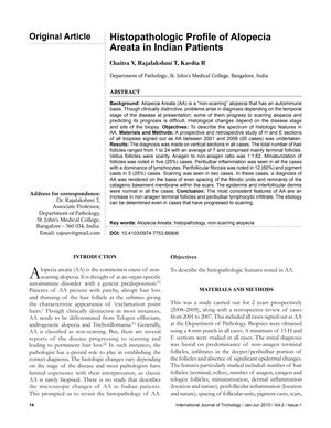
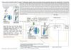
The study investigated the histopathologic characteristics of alopecia areata (AA) in 20 Indian patients, aged between 9 and 42 years, with a slight majority being female. Conducted from 2001 to 2009, it identified consistent features of AA, including increased non-anagen terminal follicles and peribulbar lymphocytic infiltrate. Additionally, 25% of cases showed follicle miniaturization, 60% had perifollicular fibrosis, and 25% exhibited pigment casts. Notably, scarring, atypical for AA, was observed in two cases. The research concluded that the etiology of AA, even in scarred cases, can be determined histopathologically, and that vertical sections are adequate for diagnosis, allowing pathologists to confidently identify AA based on these features.
 MagicBold