Investigation of Hair Shaft in Seborrheic Dermatitis Using Atomic Force Microscopy
January 2011
in “
Skin Research and Technology
”
TLDR Hair from people with seborrheic dermatitis is thicker scaled, more damaged, and thinner than healthy hair, and atomic force microscopy can help monitor the condition.
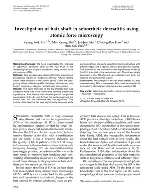
The study analyzed hair samples from 15 patients with seborrheic dermatitis (SD) and 15 healthy controls using atomic force microscopy (AFM). It found that SD-affected hair had a sevenfold increase in scale thickness compared to control hair, and the diameter of lesional hair was 10-35% less than perilesional hair. The cuticle of lesional hair was significantly damaged, although roughness parameters did not show significant differences. The study also noted that the diameter of SD-affected hair did not thin as much as in androgenetic alopecia and observed intra-hair shaft diameter diversity within the same hair shaft, which is typically a sign of follicular miniaturization in androgenetic alopecia. The study concluded that AFM is a useful non-invasive tool for monitoring SD treatment response and severity, and that hair shaft physical properties can help differentiate between inflammatory scalp diseases. Further research is needed to understand the relationship between hair density and diameter in SD-affected hair.