Dermoscopy and Trichoscopy in Dermatomyositis: A Cross-Sectional Study
January 2022
in “
Journal of Clinical Medicine
”

TLDR Dermoscopy and trichoscopy are useful for diagnosing skin signs in Dermatomyositis.
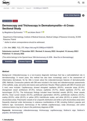
A study conducted from November 2020 to October 2021 involving 15 patients with Dermatomyositis (DM) used dermoscopy and trichoscopy to analyze skin manifestations of the disease. The most common findings in nailfold capillaroscopy were elongated capillaries (90.9%), avascular areas (81.8%), and disorganized vessel architecture (81.8%). Trichoscopy of the scalp showed yellow dots and hair diameter diversity in 60.0% of cases. Dermoscopy of Gottron’s Papules showed dotted vessels in 92.3% of cases. The study concluded that these techniques can be useful non-invasive diagnostic tools for assessing skin manifestations in DM. However, the study's limitations include a small sample size, single-center design, inclusion of only Caucasian patients, and lack of histopathological correlation. Further research is needed to evaluate the applicability of these methods in the differential diagnosis of DM.


