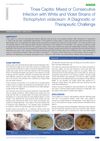
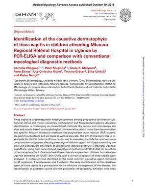
Identification of the Causative Dermatophyte of Tinea Capitis in Children Attending Mbarara Regional Referral Hospital in Uganda by PCR-ELISA and Comparison with Conventional Mycological Diagnostic Methods
October 2016
in “
Medical mycology
”
tinea capitis dermatophytes Trichophyton violaceum Microsporum audouinii Trichophyton soudanense Trichophyton rubrum PCR-ELISA mycological diagnostic methods systemic therapy topical treatments endothrix hair follicle invasion ringworm of the scalp fungal infection molecular technique systemic treatment topical treatment
TLDR PCR-ELISA is better for identifying the fungus causing scalp infections in Ugandan children than traditional methods.
In a study conducted at Mbarara University of Science and Technology in Uganda, 115 children aged 1 to 16 years with a clinical diagnosis of tinea capitis were examined to identify the causative dermatophytes of the infection. The study compared conventional mycological methods with PCR-ELISA, a molecular technique, for the detection of dermatophyte DNA. The results showed that Trichophyton violaceum was the most prevalent pathogen, followed by Microsporum audouinii, T. soudanense, and T. rubrum. The findings emphasized the importance of early and accurate identification of the dermatophyte responsible for tinea capitis to effectively manage the disease, trace its source, and prevent its spread. The study suggested that systemic therapy should be preferred over topical treatments for children in Western Uganda due to the endothrix nature of the hair follicle invasion by the most common dermatophyte, T. violaceum.