Automated Skin Surface Phenotype for Melanoma Risk Assessment
April 2023
in “
Journal of Investigative Dermatology
”

TLDR An automated method accurately assesses melanoma risk using 3D body images to analyze skin traits.
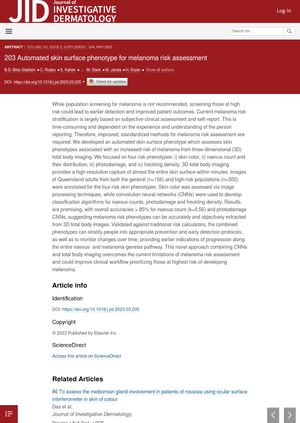
The document discusses the development of an automated skin surface phenotype for melanoma risk assessment. This method uses three-dimensional (3D) total body imaging to assess four risk phenotypes associated with an increased risk of melanoma: skin color, naevus count and their distribution, photodamage, and freckling density. The study used images of 156 Queensland adults from the general population and 300 from high-risk populations. Skin color was assessed via image processing techniques, while convolution neural networks (CNNs) were used to develop classification algorithms for naevus counts, photodamage, and freckling density. The results showed overall accuracies greater than 85% for naevus count and photodamage CNNs, suggesting that melanoma risk phenotypes can be accurately and objectively extracted from 3D total body images. This approach could improve clinical workflow by prioritizing those at highest risk of developing melanoma.


