Normolipemic Xanthoma Associated With Folliculotropic Mycosis Fungoides
May 2024
in “
Australasian journal of dermatology
”
folliculotropic mycosis fungoides alopecia xanthoma atypical T cells follicular epithelium cyst formation follicular mucinosis histiocyte infiltration lipoproteins local radiotherapy narrow-band ultraviolet B systemic etretinate intravenous interferon-gamma fMF hair loss skin masses UVB therapy etretinate interferon-gamma
Studysummary This study reports on a unique clinical presentation of folliculotropic mycosis fungoides in a 60-year-old man, highlighting complexities in diagnosis due to dense histiocytic infiltration, which led to xanthoma formation before treatment, and obscured lymphomatous features. Our plain-language summary of this paper — not a Tressless recommendation.
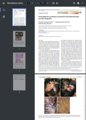
A 60-year-old Japanese man with a 4-year history of multiple yellowish masses and patchy alopecia on the scalp was diagnosed with folliculotropic mycosis fungoides (fMF), a variant of mycosis fungoides, and xanthoma. fMF is characterized by the infiltration of atypical T cells into the follicular epithelium, leading to cyst formation, alopecia, and follicular mucinosis. The patient's condition was complicated by the presence of xanthoma, a rare occurrence in fMF. The study suggests that the tumor microenvironment first triggers histiocyte infiltration around the follicles, and then these histiocytes may phagocytose lipoproteins released from destroyed follicular units, leading to xanthoma formation. The patient's treatment, which included local radiotherapy, narrow-band ultraviolet B, systemic etretinate, and intravenous interferon-gamma, helped reduce the scalp masses, leaving xanthoma.