Lupus Nephritis with Visual Field Defect Secondary to Hypertensive Retinopathy: A Case Report
November 2017
in “
Journal of Surgical Academia
”
New to Prednisolone? There is a guide in the encyclopedia. Read the guide → 
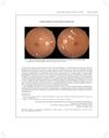
Studysummary This case report describes a woman with systemic lupus erythematosus who developed non-progressive visual field defects after hypertensive retinopathy, with cotton wool spots indicating retinal nerve fiber microinfarctions.
Automatically generated from the study's abstract, not written by a person, and not a review of the full paper. Not medical advice or a treatment recommendation. Read the original study, and consult a qualified healthcare professional before changing treatment. Full disclaimer