Erosive Pustular Dermatosis of the Scalp: Clinicopathological Correlation Leading to a Definition of Diagnostic Criteria
June 2021
in “
Wounds-a Compendium of Clinical Research and Practice
”

TLDR Chronic scalp lesions with crusts and pus that heal with strong topical steroids suggest Erosive Pustular Dermatosis, confirmed by biopsy showing specific immune cells.
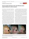
The study examined Erosive Pustular Dermatosis of the Scalp (EPDS) in 21 elderly patients, finding it common in those with androgenetic alopecia and field cancerization of the capillitium. EPDS was often triggered by treatments for actinic keratosis lesions, photodynamic therapy, cryotherapy, trauma, and surgery. It presented as crusts and pus over shiny granulation tissue, with histopathological findings showing an ulcerated epidermis and dermal infiltrates dominated by lymphocytes and plasma cells. Plasma cells were found in all biopsies and were a common criterion for diagnosis. The erosive lesions healed well within weeks after therapy with topical steroids. The study concluded that chronic, poorly healing lesions with crusts and pus over shiny granulation tissue on the scalp are suggestive of EPDS, confirmed by biopsy. Histological clues include dermal infiltrates of plasma cells and lymphocytes. Topical application of high-potency steroids was effective in treatment.


