TLDR The glassy layer of hair follicles has different fibril sizes and arrangements in guinea pigs and young mice.
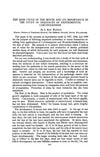
The study examines the glassy layer of hair follicles in young mice and adult guinea pigs using an electron microscope. In guinea pigs, the glassy layer consists of collagen-type fibrils about 500 Å in diameter, organized into two arrays: an inner array parallel to the follicle length and an outer array at right angles. In young mice, the fibrils are about 200 Å in diameter and develop between the outer root sheath and a surrounding layer of fibroblasts.
 521 citations
,
January 1954 in “Physiological Reviews”
521 citations
,
January 1954 in “Physiological Reviews” Hair growth is cyclic and influenced mainly by local factors.
 111 citations
,
March 1951 in “Annals of the New York Academy of Sciences”
111 citations
,
March 1951 in “Annals of the New York Academy of Sciences” Understanding the mouse hair cycle is crucial for cancer research.
April 2019 in “The journal of investigative dermatology/Journal of investigative dermatology” A specific mutation in the TRPV3 gene causes hair follicle cells to develop improperly, leading to hair loss.
10 citations
,
October 2000 in “PubMed” E6/E7 oncogenes in hair follicles cause continuous hair growth by skipping the resting phase.
122 citations
,
July 1994 in “Journal of Investigative Dermatology”  94 citations
,
February 1994 in “The journal of investigative dermatology/Journal of investigative dermatology”
94 citations
,
February 1994 in “The journal of investigative dermatology/Journal of investigative dermatology” EGF makes hair follicles grow longer but stops hair production.
59 citations
,
August 1981 in “PubMed” Trichilemmal keratinization is a unique process in hair follicles where the outer root sheath turns into keratin without a specific layer.