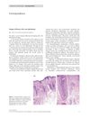
Dermoscopy Before and After Treatment of Cutaneous Larva Migrans: Through the Dermoscope
January 2022
in “
Indian dermatology online journal
”
TLDR Dermoscopy may not show hookworms clearly, and comparing it with tissue studies could improve diagnosis accuracy for skin conditions caused by parasites.
The document discusses a case of Cutaneous larva migrans (CLM), a skin condition caused by the larvae of certain parasites, in an adult male. The patient, a paddy farmer, presented with an itchy, exudative eruption on both feet. Dermoscopic examination was carried out before and after treatment, revealing various features such as a pink and purple structureless area in a linear, serpiginous, and winding pattern, corresponding to the track created by the migration of the larva. After treatment, the dermoscopic findings showed changes such as golden-brown to dark brown and black crust against a pale brown structureless background arranged in a linear serpiginous track-like fashion. The document concludes that the typical magnification offered by dermoscopy (10–20 ×) may not be enough to discern a hookworm, one of the causes of CLM, and suggests that histopathological correlation with dermoscopic findings may further elucidate the accuracy of dermoscopy in the diagnosis of this entity.


