Dermal Mass Aspirate from a Persian Cat
December 2003
in “
Veterinary clinical pathology
”

TLDR The Persian cat has a skin infection caused by a fungus, treatable with antifungal medication.
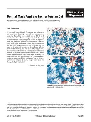
A 1-year-old spayed female Persian cat presented with alopecia, weight loss, and multiple ulcerated dermal nodules. Cytologic examination of a nodule aspirate indicated pyogranulomatous inflammation, septate hyphae, and arthrospores, leading to a diagnosis of dermatophytic pseudomycetoma. Histological analysis confirmed the presence of granulomas, septate hyphae with spores, and a Splendore-Hoeppli reaction, with a histologic diagnosis of pseudomycetoma-associated chronic multifocal severe granulomatous dermatitis. The fungus Microsporum canis was cultured from the lesion. Pseudomycetomas, common in Persian cats due to a possible heritable predisposition, are characterized by multiple lesions, absence of prior skin trauma, association with dermatophytes (especially Microsporum canis), and a distinct histological appearance compared to fungal mycetomas and dermatophytosis. The prognosis for pseudomycetoma is fair with systemic antifungal treatment, and such a diagnosis should be considered in Persian cats with similar symptoms and cytologic findings.


