Diagnosis and Treatment of Alopecia in an Older Dog with Abdominal Mass
November 1998
in “
Journal of Small Animal Practice
”
alopecia telogenization hyperadrenocorticism adrenal tumor sex-hormone disorder testicular neoplasia telogen effluvium hypothyroidism follicular dysplasias epitheliotrophic lymphoma necrolytic migratory erythema demodicosis dermatophytosis Sertoli cell tumor preputial erythema radiography ultrasonography histopathology hair loss hair shedding Cushing's disease adrenal gland tumor hormone imbalance testicular cancer stress-induced hair loss underactive thyroid skin lymphoma skin redness X-ray ultrasound tissue examination
TLDR The dog had a Sertoli cell tumor, which was successfully removed with surgery.
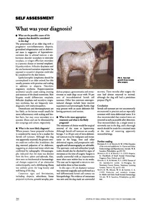
The document discusses the diagnosis and treatment of alopecia in an older dog presenting with non-inflammatory alopecia, generalized telogenization, and an abdominal mass. The possible causes considered included hyperadrenocorticism due to an adrenal tumor, a sex-hormone disorder secondary to testicular neoplasia, or telogen effluvium secondary to systemic disease or internal neoplasm. Other conditions such as hypothyroidism, follicular dysplasias, epitheliotrophic lymphoma, necrolytic migratory erythema, demodicosis, and dermatophytosis were also considered. The most likely diagnosis was a Sertoli cell tumor, indicated by linear preputial erythema and confirmed by radiography and ultrasonography. The treatment of choice was surgical removal of the mass, which was a 10 cm diameter tumor confirmed as a well-differentiated Sertoli cell tumor on histopathology. The dog recovered uneventfully, with coat returning to normal within three to four months post-surgery. The document concludes by recommending the early removal of retained testes to prevent Sertoli cell tumors, which are not uncommon in dogs with intra-abdominal testes.
