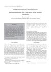
Association of Systemic Involvement with Skin Morphology Assessed by Multiphoton Microscopy in Pseudoxanthoma Elasticum
October 2021
in “
Journal of Investigative Dermatology
”
Pseudoxanthoma elasticum PXE ABCC6 gene multiphoton microscopy calcification skin morphology Phenodex score Skin-score Eye-score calcification area calcification density redundant skin folds inflammatory skin signs internal carotid artery hypoplasia aortic calcification peripheral atherosclerosis Vascular scores MPM
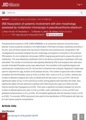
TLDR Skin changes in Pseudoxanthoma elasticum patients can indicate the severity of related health issues.
The study investigated the association between skin morphology and systemic involvement in 25 patients with Pseudoxanthoma elasticum (PXE), a connective tissue disorder caused by mutations in the ABCC6 gene. The researchers used multiphoton microscopic (MPM) imaging to assess the extent of calcification in the skin. They found a correlation between the number of typical skin sites affected by PXE and the Phenodex score (ρ=0.55, p=0.004), Skin- score (r=0.75, p=<0.001), and the number of affected non-typical skin sites correlated with the Eye-score (r=0.4, p=0.047). Higher calcification area (CA) (p= 0.012) and calcification density (CD) (p=0.032) were found in patients with redundant skin folds. Higher CD was also observed in patients with inflammatory skin signs (p=0.039) and with internal carotid artery hypoplasia (p=0.045). There was a significant correlation between CA and the number of affected typical skin sites (r=0.44, p=0.026), aortic calcification (r=-0.44, p=0.027) and peripheral atherosclerosis (r=0.4, p=0.02). CD correlated significantly with the Vascular scores (r=0.45, p=0.022). The study concluded that MPM could be useful for identifying PXE patients at high risk for more severe systemic complications.