Application of 3D Reflectance Confocal Microscopy: Melanocytic Proliferations as 3D Models; JAAD Supplemental Material
August 2020
TLDR 3D models from confocal microscopy improve melanoma detection on sun-damaged skin.
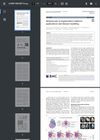
The study explored the use of 3D reflectance confocal microscopy (RCM) to improve the detection of melanoma on chronically sun-damaged skin by creating 3D models from 2D confocal image stacks. The research demonstrated that 3D reconstructions could provide enhanced visualization of melanocytic proliferations, revealing details such as pleomorphism, cell uniformity, and the distribution of melanocytic cells within the skin layers. The process involved acquiring confocal images with the Vivascope 1500, aligning and preprocessing them in ImageJ, and using TrakEm2 for manual segmentation and reconstruction. The study highlighted the potential of 3D models to expand the data available for diagnosing skin conditions, offering a more comprehensive view of skin biology compared to traditional 2D methods.